
Figure 6. Synergistic anti-proliferating effect of DCA in combination with doxorubicin in primary leukemic cells.
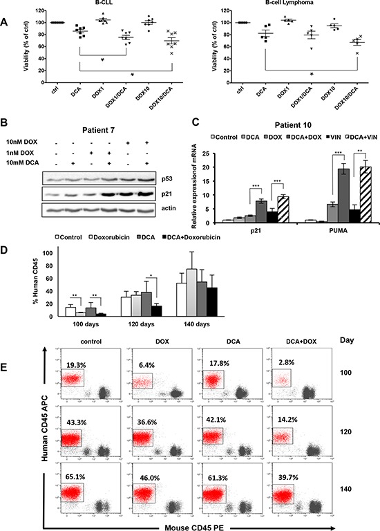
A. Tumor cells from seven patients with B-CLL and five patients with B-cell lymphoma (all wt p53) were incubated with 10 mM DCA alone or in combination with doxorubicin (1 or 10nM; DOX) for 72 h. Tumor cells were identified (CD19+/CD20+) by flow cytometry and tumor cell death was quantified by 7-AAD staining using the Muse® Cell analyzer (values are the mean ± SEM from one experiment performed in triplicate *p < 0.05). B. Tumor cells (wt p53) from a patient with B-CLL (Patient 7) were treated with 10 mM DCA alone or in combination with doxorubicin (DOX) for 24 h. p53 and p21 expression was assessed by western blotting. C. mRNA levels of p21 and PUMA in primary tumor cells from a patient with MDS (wt p53; Patient 10) following incubation or not with 10 mM DCA for 24 h and then addition or not of 10 nM doxorubicin (DOX) or 10 nM vincristine (VIN) for another 24 h before analysis by RT-qPCR; **p < 0.01; ***p < 0.001. See also Supplementary Figure S6 (values are the mean ± SEM from one experiment performed in triplicate). D. NSG mice were engrafted with primary human AML cells (wt p53). At day 80 post-graft, they were treated with doxorubicin (n = 4), DCA (n = 4) or both (n = 4) and the percentage of human cells in peripheral blood samples was measured every 20 days; *p < 0.05; **p < 0.01 (values are expressed as median ± SEM). E. Representative plots of the percentage of human tumor cells in peripheral blood samples of mice at day 100, 120 and 140 post-graft.