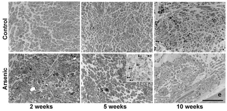
Figure 2.
Arsenic induced changes in microbial community structure as observed by TEM at 2, 5, and 10 weeks (top row – control, bottom row - arsenic exposed), (s) spore. Insert shows abundance of spores (arrows) in 5 week arsenic exposed mice, light micrograph. The absence of the small coccoids and presence of filamentous bacteria in the mucosa closest to the epithelium is most pronounced in the 10 week arsenic exposed mice. Bars 5 μm.

