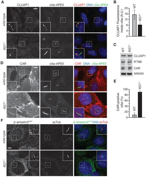
Figure 7. Validation of CLUAP1 depletion and CAR and β-arrestin 2 enrichment in Ift27−/− cilia.
(A) Wild-type and Ift27−/− cells expressing cilia-APEX were starved for 16 h to induce ciliation, fixed in 4% PFA, permeabilized using 0.1% saponin and stained for endogenous CLUAP1. Merged insets show primary cilia with channels shifted to aid visualization. Yellow arrows point at the basal body.
(B) Ciliary CLUAP1 signals in cilia were quantified in wild-type (n = 52) and Ift27−/−(n = 42). Error bars depict standard error of the mean.
(C) Western blots of wild-type and Ift27−/− cell lysates.
(D) Immunofluorescence microscopy as in (A) using anti-CAR antibody to stain native CAR.
(E) Number of CAR-positive (CAR+) and CAR-negative (CAR−) cilia were counted in indicated cell lines and displayed as bar graphs. Error bars depict standard deviations (n = 3).
(F) Wild-type and Ift27−/− cells stably expressing β-arrestin2-GFP were analyzed by fluorescent microscopy as in (A). All scale bars are 5 μm (main panels) and 1 μm (insets). See also Figure S5.

