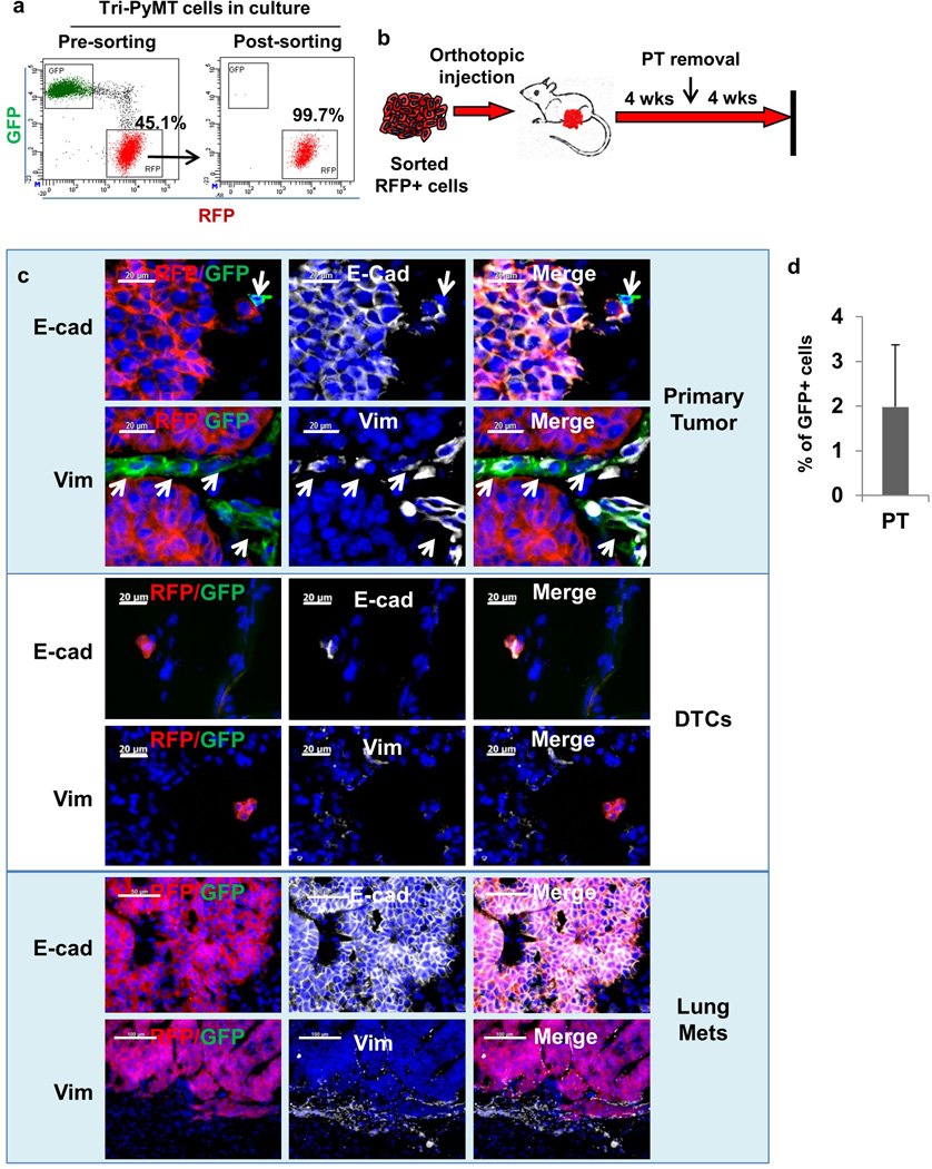
Extended Data Figure 5. Establishing an orthotopic model with sorted RFP+ Tri-PyMT cells.
a, Flow cytometry plots show Tri-PyMT cells before and after sorting for RFP+ cells. Numbers indicate the percentage and purity of RFP+ cells used for establishing orthotopic breast tumors in mice.
b, Schematic of the orthotopic breast tumor model with sorted RFP+ Tri-PyMT cells. Cells are injected into the mammary gland of wild type mice to generate primary breast tumors, resection of primary tumor at 4 weeks and lung metastases evaluation in another 4 weeks.
c, Characterization of tumor cells in the primary tumor, disseminated tumor cells (DTCs) and tumor cells in the lung metastasis of the Tri-PyMT orthotopic model. Sections of primary tumors and lungs from Tri-PyMT orthotopic mice were immunostained for E-cadherin and Vimentin (in white pseudocolor). Essentially all RFP+ tumor cells are detected as E-cad+/Vim−, while the scattered GFP+ tumor cells in the primary tumor are E-Cad−/Vim+ (as indicated by arrows in the top panel). Representative images are shown (n=8). d, Plot shows the percentage of GFP+ cells out of total tumor cells (GFP+ plus RFP+, n=6).

