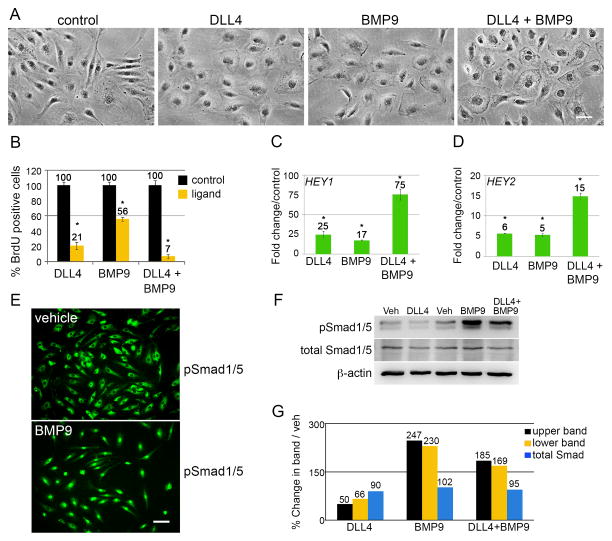
Figure 1. DLL4/Notch and BMP9 cooperatively suppress endothelial cell proliferation and induce HEY genes.
A) Primary human aortic endothelial cells (HAEC) were stimulated with ligand as described for 24h. Scale bar = 50μm. B) After 18h treatment, cells were given 10μg/mL BrdU for the last 6h of growth. Cells were fixed at 24h and processed for immunostaining with anti-BrdU. The percentage of BrdU positive cells was calculated (n=10 per condition). Graphed are means ±SD. C–D) Cells under the same experimental conditions were grown for 24h, and total RNA was collected for qPCR. Primers were used to amplify HEY1 and HEY2 transcripts normalized to PPIA (cyclophilin). Graphed are fold change means compared to control ±SD. E) HAEC were synchronized by serum starvation for 8h, treated with vehicle or BMP9 for an additional 16h, then fixed and stained for phospho-Smad1/5 (pS463/465). Scale bar = 50μm. F) HAEC were treated with vehicle or ligands for 24h and whole cell lysates were immunoblotted for phospho-Smad1/5 (pSmad1/5) and total Smad1/5 (total Smad1/5). pSmad1/5 present as a doublet and the bands were quantified separately. G) Quantified immunoblot bands were normalized to β-actin and represented as a percentage of vehicle. (*) indicates a statistical significance with p<0.05.

