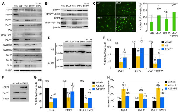
Figure 3. P27KIP1 mediates the quiescence signal of DLL4 and BMP9.
Primary human aortic endothelial cells (HAEC) were treated with ligands as indicated. A–B) Cell lysates were collected after 24h for immunoblot using antibodies against cell cycle regulatory proteins. Quantification of the immunoblot bands in panels A, B and E are in Supplementary Table I. C) Cells stimulated under the same conditions were fixed after 24h and immunostained for P27KIP1 (scale bar=100μm), and nuclear fluorescence intensity was quantified. Graphed are means relative to vehicle ±SD, N≥5 fields per condition. D) P27KIP1 was targeted using siRNA (siP27) compared to non-targeting (NT) siRNA, and protein levels detected by immunoblot. E) Proliferation following knockdown of P27KIP1 was measured by BrdU incorporation. F) Cells were transduced with adenoviral vectors with control LacZ (adLacZ) or an expression construct for SKP2 (adSKP2). Cell lysates were immunoblotted, validating the overexpression of SKP2 (2232% of control) and consequent downregulation of its target P27KIP1 (63% of control). G) Cells transduced with control AdLacZ or AdSKP2 were stimulated with ligands or vehicle control, and proliferation measured by BrdU. SKP2 overexpression reversed the proliferation effects of the ligands (G) and significantly decreased the ligand-mediated nuclear intensity of P27KIP1 (H). Graphed are means ±SD. (*) signifies statistical significance of P<0.05. (†) signifies statistically significant reversal of effects (p<0.05).

