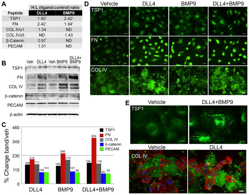
Figure 4. Extracellular matrix proteins accumulate following DLL4 and BMP9 stimulation.
A) Primary human aortic endothelial cells (HAEC) were stimulated with ligands as described. Proteins were collected and conjugated to isotope coded affinity tags and analyzed by mass spectrometry. Heavy (H) to light (L) isotope ratios of specific peptides reflected fold changes in proteins in ligand-stimulated conditions to control conditions. ND indicates peptides not detected. (*/†) indicate peptides detected in multiple cellular fractions. B) Cell lysates were collected 24h after ligand treatment, and immunoblotted to independently validate mass spectrometry analysis. C) Quantified immunoblot bands are represented as a percentage of vehicle. D) Cells were fixed and processed for immunofluorescence staining to detect thrombospondin-1 (TSP1), fibronectin (FN), or collagen type IV (COL IV) under the conditions indicated. Scale bar = 50μm. E) Representative high magnification views are shown to demonstrate the fibrillar, cell surface and extracellular accumulation of TSP1 and COL IV. All ECM proteins are shown in green, phalloidin-stained actin filaments are red, and DAPI-stained nuclei are blue. Scale bar = 30μm for the TSP1 panels and 50μm for the COL IV panels.

