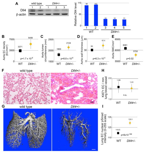
Figure 6. Haploinsufficiency of DLL4 increases aortic endothelial density, and increases phospho-Smad1/5 levels in lung endothelial cells of adult mice.
A) Protein lysates collected from thoracic aortas of Dll4+/− mice and wild type (WT) littermates were immunoblotted to determine levels of DLL4. Quantified immunoblot bands are represented as a percentage of Dll4+/− relative to the average of the WT bands. B) Thoracic aortae were fixed, stained with Hoechst nuclear dye and endothelial density measured by immunofluorescent imaging. Graphed are means ± SEM. C–E) Transverse sections of paraffin embedded thoracic aortas of littermates were H&E stained, and lumen circumference (C), aorta wall thickness (D), and smooth muscle cell density (E) were measured. N=9 mice/group. Graphed are means ± SEM. F) Sections of paraffin embedded lungs from littermates were H&E stained, revealing extensive red blood cell infiltration of alveolar spaces. Scale bar = 50μm. G) Lung vasculature was perfused with radiopaque silicone rubber (Microfil), and imaged by microcomputed tomography. Irregular vascular branching patterns can be observed in Dll4+/− lung. Scale bar = 1mm. H) Sections of paraffin embedded lungs were immunohistochemically stained for the ki67 proliferation marker, and endothelial cells positive for the marker were counted, and quantified relative to total vessel lumen circumference. Graphed are means ± SEM. N=7 mice/group. I) Sections of paraffin embedded lungs were immunohistochemically stained for phospho-Smad1/5, and staining intensity of endothelial nuclei was measured. Graphed are means ± SEM. N = 7 Dll4+/− mice (31,195 nuclei) and 8 WT mice (19,101 nuclei).

