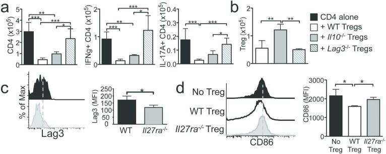
Figure 6. Lag3 dependent inhibition of T cell activation in the draining lymphoid tissues.
(a) Rag1 −/− mice received CD45.1+ CD4 T cells alone, or together with WT, Il10 −/− , or Lag3 −/− Tregs. T cell expansion and cytokine expression in the mLN were determined 7 days post transfer. (b) Number of Tregs in the mLN. (c) Lag3 expression on WT and Il27ra −/− Tregs. (d) mLN DC (CD11c+ MHCII+) expression of CD86 was determined at 4 days post T cell transfer. Data shown represent the mean ± SD of 3~8 individually tested recipients. *, p<0.05; ***, p<0.001.

