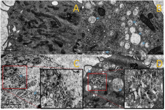
Fig. 9.

TEM images of DCs after LPK NPs internalization. (A) Control group, (B) LPK− NPs, (C) LPK+ NPs, (D) LPKpH NPs. Blue arrows show the position of endosomes, which were more numerous in groups treated with NPs compared to the control group, and LPKpH NPs underwent significant degradation in endosomes. Images in black boxes are zoomed-in pictures of endosomes. (For interpretation of the references to colour in this figure legend, the reader is referred to the web version of this article.)
