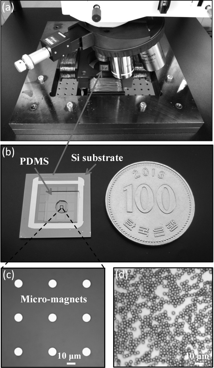
FIG. 1.
Experimental setup. (a) Four solenoid coil magnets produce the uniform in-plane field. The studied sample is placed at the center of the field area and below the optical microscope. The field strength is monitored from the Gaussmeter positioned below the sample. (b) Micro-magnet arrays deposited on a Si wafer and the Polydimethylsiloxane (PDMS) well for localizing the experimental solution. (c) Microscopic image of the micro-magnet array. (d) Optical image of Dynabeads M-280 Streptavidin beads (2.8-μm diameter).

