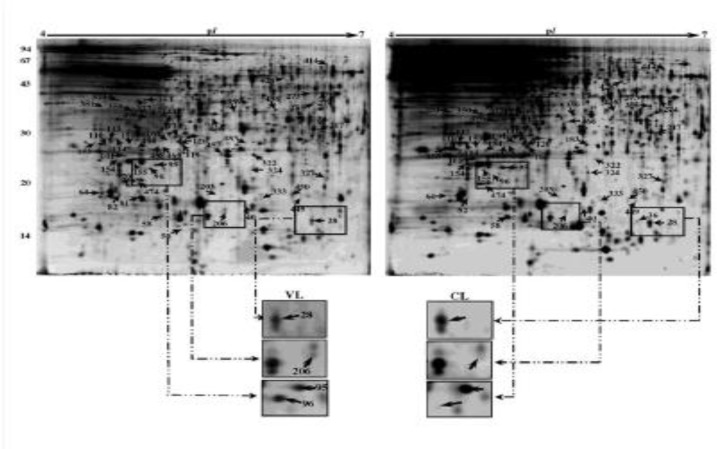
Fig. 1.
2-D gel analysis of proteins extracted from Leishmania tropica isolated from cutaneous and visceral (spleen) of a 5-month old dog. In first dimension (IEF), 120 µg was loaded on a 18-cm IPG strip with a linear gradient of pH 4-7. In the second dimension, 12% SDS-PAGE gels were used, with a well for molecular weight standards. Proteins were visualized by silver staining. Arrows represent spots identified by MS (Tables 2-3). Examples of changes in protein abundance between viscerotropic (VTI) and cutaneous (CI) samples have been presented

