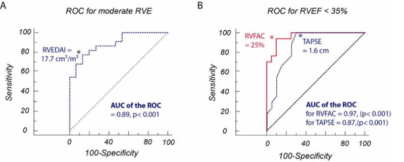
Figure 5.

Receiver operating characteristic curves: (A) Detecting RV dilatation (MRI RVEDV index > 104 mL/m2 for women and > 115 mL/m2 for men) by echocardiographic RVEDA index (area under curve 0.89). (B) Detecting moderately diminished RV systolic function (MRI RVEF < 35%) by echocardiographic RVFAC (area under curve 0.97) and TAPSE (area under curve 0.87). P = 0.07 for comparison between curves.
