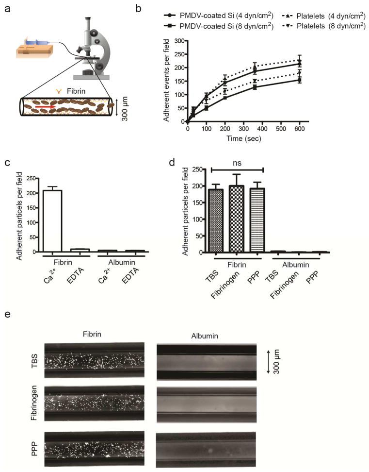
Figure 5. Adhesion of PMDV-coated Si particles to immobilized fibrin under flow.
(a) Schematics of a microtube with the inner surface coated with fibrins to simulate blood clotting. 106/ml PMDV-coated Si particles were perfused through microtubes at 4 dyn/cm2 via a controlled syringe pump. (b) Adhesion of coated particles and activated platelets to a fibrin-functionalized surface under flow. (c) Comparison of adherent particles in Ca2+- or EDTA-containing buffer under flow. For negative control, albumin was coated on the surface in the absence of fibrin. (d) Comparison of particle adhesion to fibrin-coated microtubes under flow in tris-buffered saline (TBS), TBS containing 2 mg/ml soluble fibrinogen or platelet poor plasma (PPP). TBS buffer was supplemented with Ca2+. Results are presented as the mean ± SEM; ns indicates no significant difference. (e) Representative fluorescent images of adherent particles in fibrin-coated microtubes were taken after removing unbound particles via TBS washing.

