
Figure 3.
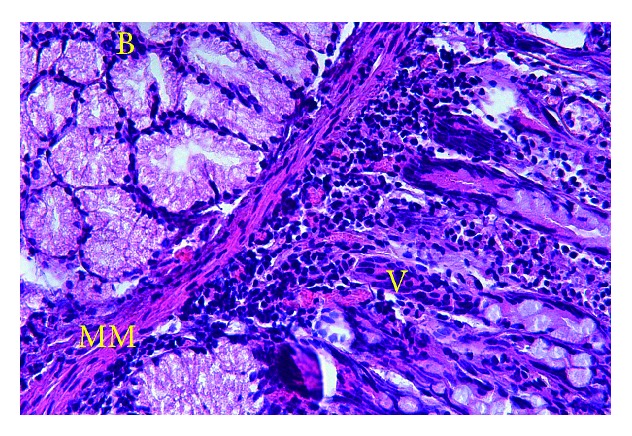
Histological view of the cyst wall showing typical structure of duodenal mucosa (hematoxylin and eosin, ×100) (B: Brunner's glands, MM: mucosa muscularis, V: duodenal villi).
Official websites use .gov
A
.gov website belongs to an official
government organization in the United States.
Secure .gov websites use HTTPS
A lock (
) or https:// means you've safely
connected to the .gov website. Share sensitive
information only on official, secure websites.
Histological view of the cyst wall showing typical structure of duodenal mucosa (hematoxylin and eosin, ×100) (B: Brunner's glands, MM: mucosa muscularis, V: duodenal villi).