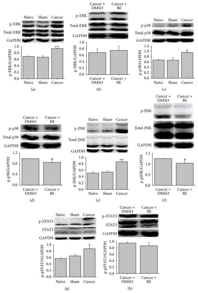
Figure 3.
Intrathecal BE inhibits the expression of p-p38 and p-JNK in spinal cord of CIBP rats. ((a), (b)) Western blot and the quantitative data of p-ERK protein in spinal cord of rats. Protein level of p-ERK significantly increased in CIBP rats compared to naïve rats. However, intrathecal BE (100 μg) had no influence on the expression of p-ERK. ((c), (d)) Western blot and quantitative data of p-p38 protein in spinal cord of rats. Intrathecal BE (100 μg) reduced the expression of p-p38 protein in CIBP. ((e), (f)) Western blot and quantitative data of p-JNK protein in spinal cord of rats. Intrathecal BE (100 μg) reduced the expression of p-JNK in CIBP. ((g), (h)) Western blot and quantitative data of p-STAT3 protein in spinal cord of rats. Intrathecal BE (100 μg) had no influence on the expression of p-STAT3 in CIBP. Data are presented as means ± SEM (∗ p < 0.05, ∗∗ p < 0.01 versus naïve group; # p < 0.05 versus cancer + DMSO i.t. group).

