Figure 2.
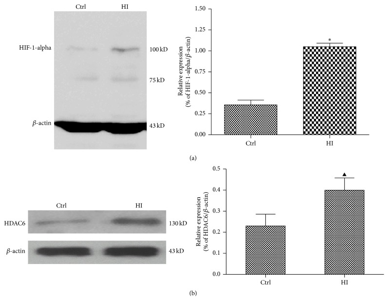
Hypoxia-ischemia induced HDAC6 expression and oxidative stress in vitro. The PC12 cells were treated with HI for 24 h. Ctrl express medium-treated group. (a) The expression of HIF-1-alpha was analyzed by Western blot. Results of the densitometric quantification are represented as mean ± SEM (n = 3). ∗ P < 0.01 versus control group. (b) The expression of HDAC6 was analyzed by Western blot. Results of the densitometric quantification are represented as mean ± SEM (n = 3). ▲ P < 0.05 versus control group.

