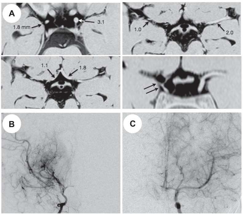
Fig. 3.
Representative three-dimensional constructive interference in steady state (A) and magnetic resonance angiography findings (B, C) of a 4-year-old girl with unilateral moyamoya disease. A: The outer diameter of the bilateral C1, M1, and A1 is printed. Note the marked shrinkage of the right carotid fork on a coronal image (double arrows). B: Right carotid angiogram shows severe stenosis of the carotid fork and marked development of basal moyamoya vessels, being graded as stage 3. C: Left carotid angiogram shows no definite abnormality (stage 0).

