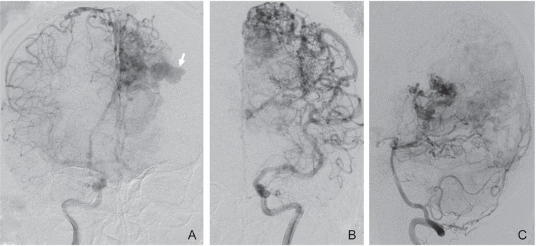
Fig. 1.
Angiograms obtained before hemorrhage. Anteroposterior view of the right internal carotid artery (A), the left internal carotid artery (B), and the left vertebral artery (C) angiogram showing a left parieto-occipital arteriovenous malformation fed by numerous cortical branches with an intranidal aneurysm (arrow).

