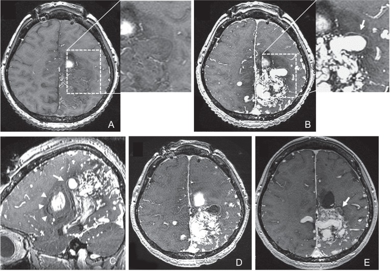
Fig. 3.
Axial non-enhanced (A) and gadolinium-enhanced (B) T1-weighted image obtained on day 2 revealing the linear enhancement of the outer surface of the thickened vessel wall of the intranidal aneurysm (arrows). Sagittal gadolinium-enhanced T1-weighted image (C) showed the intranidal aneurysm located adjacent to the intracerebral hematoma (arrowheads). The outer rim of the hematoma was iso-intensity. Axial gadolinium-enhanced T1-weighted image obtained 5 days after the targeted embolization (D) showing total thrombosis of the intranidal aneurysm. Axial gadolinium-enhanced T1-weighted image obtained 10 months after the targeted embolization (E) showing decrease of the vessel wall enhancement and shrinkage of the intranidal aneurysm (arrow).

