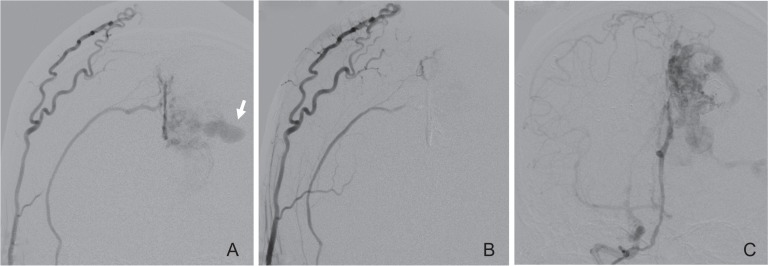
Fig. 4.
A: Right external carotid angiogram obtained before targeted embolization showing that the intranidal aneurysm (arrow) is mainly fed by the right middle meningeal artery through the falx. B: Right external carotid angiogram after the embolization showing no contrast filling of the aneurysm. C: Right internal carotid angiogram after the embolization also showing no contrast filling of the aneurysm.

