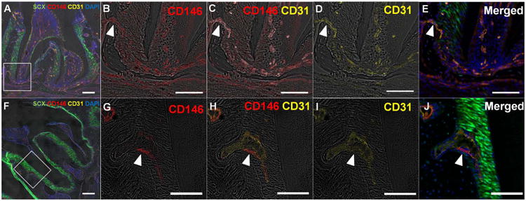
Figure 1. Co-immunofluorescent staining of CD146 with endothelial cell marker, CD31 in scxGFP mouse 2nd molar at 3 weeks (A-E) and 3 months (F-J) of age.

(A, F) CD146+ cells were detected in association with vasculature identified by CD31, through subginigival connective tissue, PDL space, dental pulp, endosteal space, and bone marrow. (B-E, G-J) Magnified view of the inserted insets in panel A and F. CD146+ cells are derived from endosteal spaces through the channels between alveolar bone and PDL (arrowhead). Scale bar; 200μm (A,F), 100μm (B-E, G-J)
