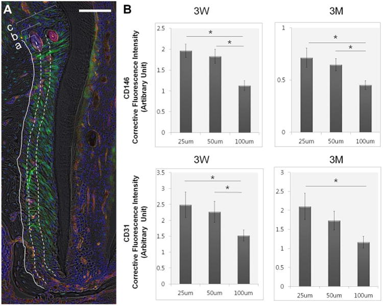
Figure 2. The distribution of CD 146+, CD31+ Cells in the vicinity of bone-PDL interface.

(A) The fluorescent intensity of CD146 and CD31 was measured within 25μm (a), 50μm (b), and 100μm (c) from B to PDL interface of mesial complex of mandibular 2nd molar, Scale bar=100μm. (B) Both CD31 and CD146 showed significantly higher fluorescence within 25μm and 50μm compared to 100μm at both ages * statistically significance (p<0.05), N=5, error bars present the standard error of the mean (SEM)
