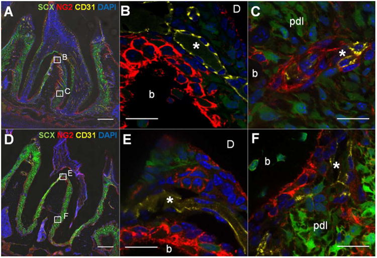
Figure 3. Co-immunofluorescent staining of NG2+ cells with endothelial cell marker, CD31 in scxGFP mouse 2nd molar at 3 weeks (A-C) and 3 months (D-F) of age.

(A,D at X20) NG2+ cells (red) were observed in proximity of CD31+ blood vessels (yellow) but also at bone-PDL and cementum-PDL interfaces. (B, E at X60) Magnified view of interradicular region. Interradicular region was intensely populated with CD31+ blood vessels (*) closely to NG2+ cell-lining bone-PDL interface. (C, F at X60) PDL adjacent to bone. NG2+ cells are located adjacent to CD31+ endothelial wall (*) within PDL. b=bone, d=dentin, Scale bar; 200μm (A, D), 20μm (B,C,E and F)
