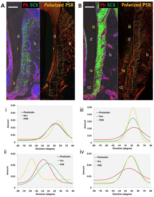
Figure 6. Orientation and organization of PDL cell cytoskeleton, fibroblasts and collagen fibers.

Immunofluorescence of phalloidin (red) and polarized light microscopy of PSR staining in scxGFP 2nd molar at 3 weeks (A) and 3 months (B). Plots illustrate directionality of birefringent collagen fibers, fibroblasts, and PDL cell cytoskeletons within mesiocoronal (i,iii) and mesioapical regions (ii,iv). Directionality of collagen fibers, fibroblasts, and PDL cell cytoskeleton coincides within PDL complex, except in apical region of 3 weeks old group. b=alveolar bone, d=dentin, cc=cellular cementum, Scale bar = 100μm
