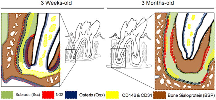
Figure 7. Schematic representation of the distribution of biomolecules identified in scxGFP mice molar at 3 weeks and 3 months.

The alveolar bone crest and periapical region are areas of high biomechanical activity and the entheses within those regions are putative locations of cellular differentiation and tissue adaptation. Perivascular niche for stemness identified using CD146 and CD31 (yellow) runs along bone-PDL entheses. NG2/Osx+ cells line the bone-PDL and cementum-PDL entheses, where biophysical signals from eruption and function are supposedly amplified. Scx are positive only in the fully developed PDL as a conveyer of biophysical cues. BSP was rich at apical bone-PDL enthesis at 3 weeks but negative at 3 months
