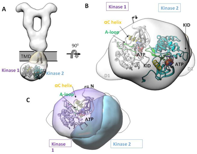
Figure 7. Asymmetric dimer of TKD.
(A) The atomic models of TM and TKD are fitted to show spatial relationships. (B) Bottom view of TKD asymmetric dimer. (C) Same view as in (B), but highlighting the catalytic site position of kinase 1, where the ATP binding pocket, the A-loop, and the αC helix are in close proximity to the neighboring kinase 2. KID = kinase insert domain. αC = control α-helix. A-loop = activation loop.

