Fig. 4.
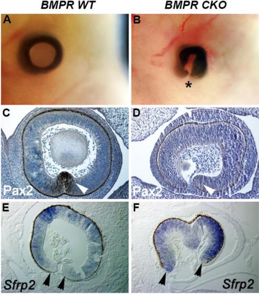
Analysis of the optic cup of E12.5 wild type eyes and eyes in which the type I BMP receptors, Bmpr1a and Acvr1 were conditionally deleted in the prospective lens placode (BMPR CKO). (A, B) The extent of coloboma formation in whole embryo heads in the BMPR CKO embryos. Asterisk indicates ventral side of tissue where fissure has not closed in the CKOs. (C, D) Immunostaining (brown staining) for Pax2 was observed at the margins of the optic fissure in wild type eyes (white arrowhead), but undetectable in the BMPR CKO eyes (white arrowhead). (E, F) Sfrp2 transcripts (blue staining) were increased at the margins of the optic fissure in BMPR CKO eyes (black arrowheads).

