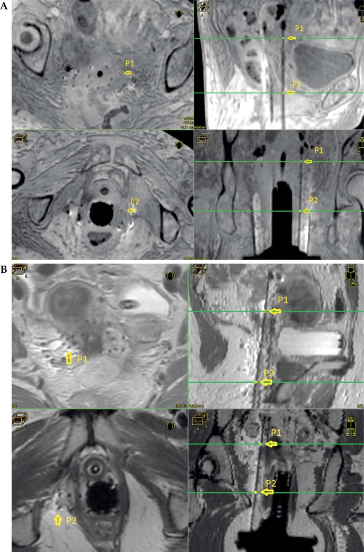
Fig. 3.
A) 3D T1W MRI images showing two different paraxial slices (left) and the choice of two points to determine the direction of one needle. Upper right and lower right show the position of points P1 and P2 in parasagittal and para-coronal views, respectively. B) T2 MRI images showing two different paraxial slices (left) and the choice of two points to determine the direction of one needle. Upper right and lower right show the position of points P1 and P2 in para-sagittal and para-coronal views, respectively

