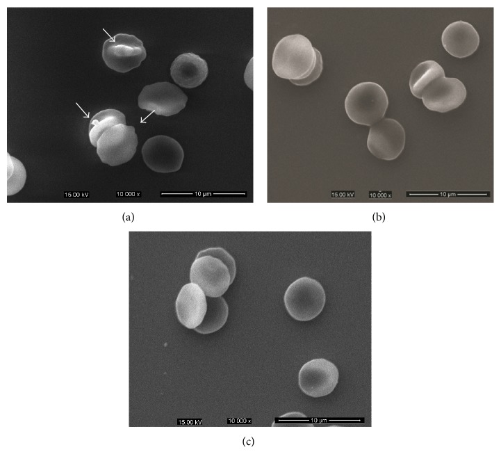
Figure 6.
Morphological changes of erythrocytes in HFD-induced and LTC-treated mice observed with scanning electron microscope. (a) Erythrocytes of HFD-induced mice deformed with protrusions or irregular appearances. (b) Erythrocytes of LTC-treated (300 mg/kg) mice presented the more typical discocytes with rare deformed cells. (c) Erythrocytes of ezetimibe-treated mice presented the more typical discocytes with rare deformed cells.

