
Figure 3.
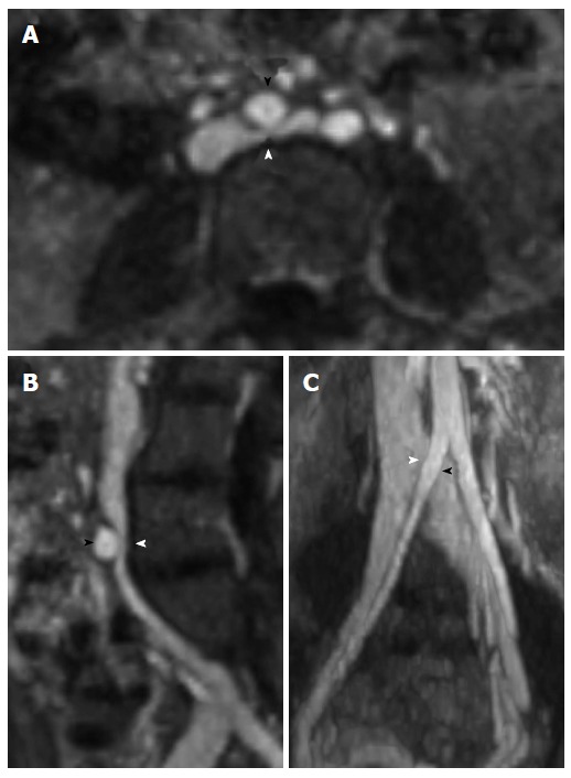
Magnetic resonance venography with axial (A), sagittal (B), and coronal (C) reformatted images demonstrating May-Thurner syndrome anatomy with compression of the left common iliac vein (white arrowhead) by the right common iliac artery (black arrowhead).