

Abstract
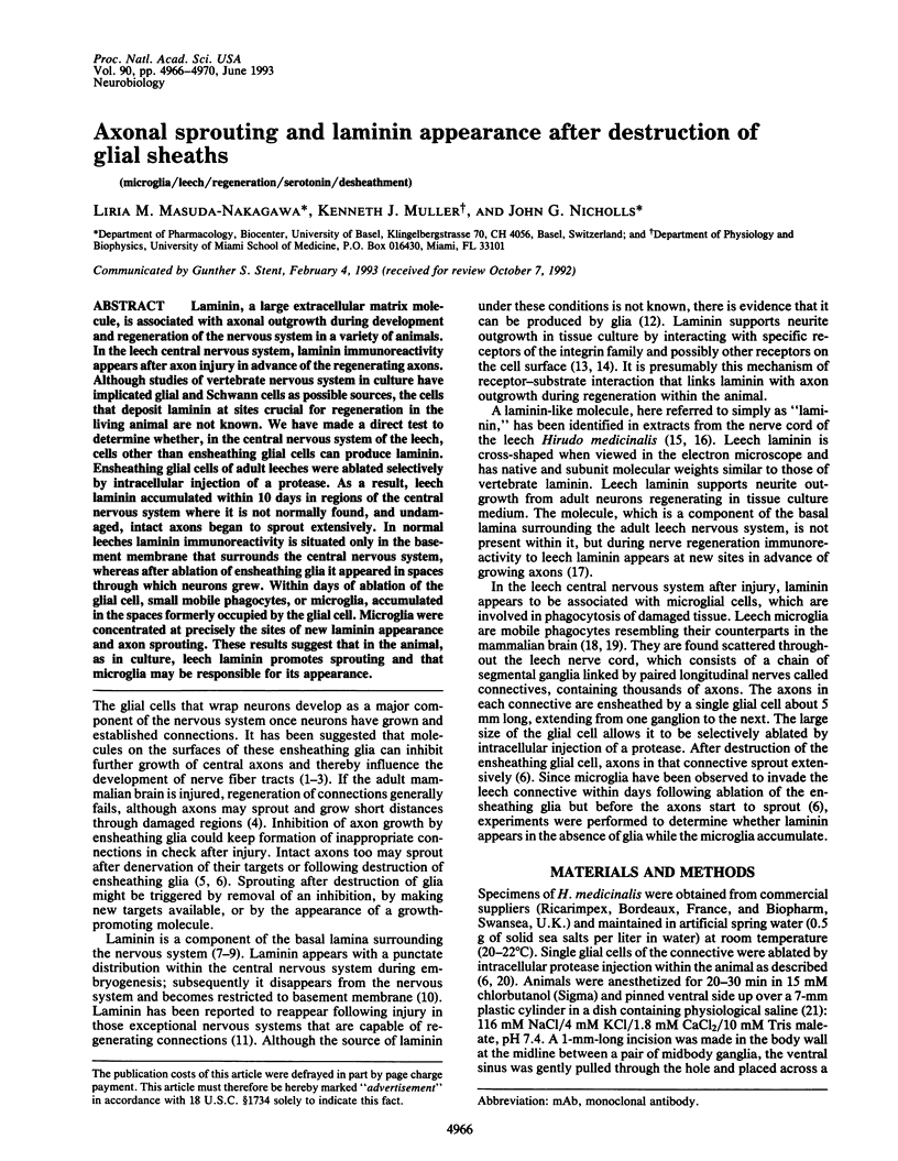
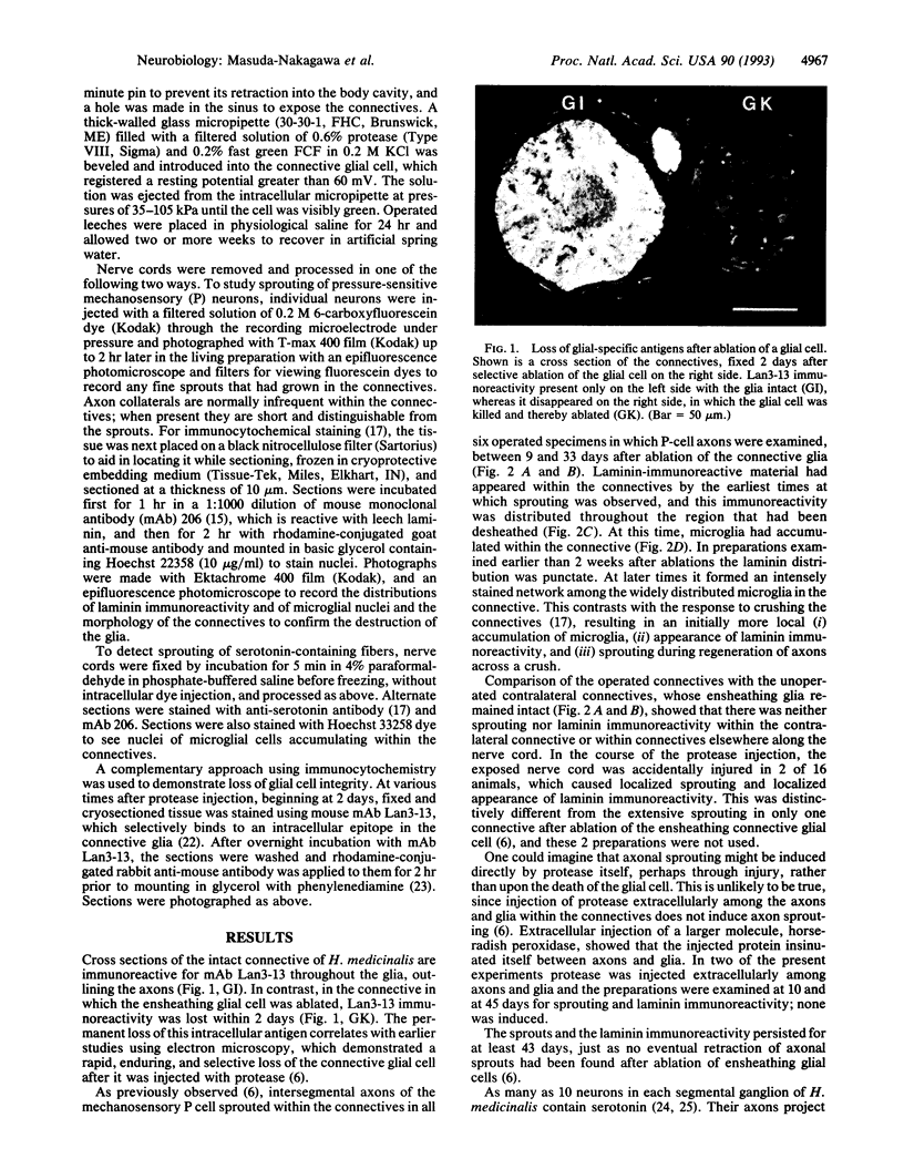
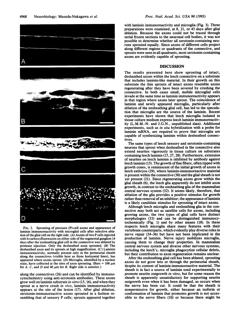
Laminin, a large extracellular matrix molecule, is associated with axonal outgrowth during development and regeneration of the nervous system in a variety of animals. In the leech central nervous system, laminin immunoreactivity appears after axon injury in advance of the regenerating axons. Although studies of vertebrate nervous system in culture have implicated glial and Schwann cells as possible sources, the cells that deposit laminin at sites crucial for regeneration in the living animal are not known. We have made a direct test to determine whether, in the central nervous system of the leech, cells other than ensheathing glial cells can produce laminin. Ensheathing glial cells of adult leeches were ablated selectively by intracellular injection of a protease. As a result, leech laminin accumulated within 10 days in regions of the central nervous system where it is not normally found, and undamaged, intact axons began to sprout extensively. In normal leeches laminin immunoreactivity is situated only in the basement membrane that surrounds the central nervous system, whereas after ablation of ensheathing glia it appeared in spaces through which neurons grew. Within days of ablation of the glial cell, small mobile phagocytes, or microglia, accumulated in the spaces formerly occupied by the glial cell. Microglia were concentrated at precisely the sites of new laminin appearance and axon sprouting. These results suggest that in the animal, as in culture, leech laminin promotes sprouting and that microglia may be responsible for its appearance.
Full text
PDF
Images in this article
Selected References
These references are in PubMed. This may not be the complete list of references from this article.
- Baier H., Bonhoeffer F. Axon guidance by gradients of a target-derived component. Science. 1992 Jan 24;255(5043):472–475. doi: 10.1126/science.1734526. [DOI] [PubMed] [Google Scholar]
- Bandtlow C., Zachleder T., Schwab M. E. Oligodendrocytes arrest neurite growth by contact inhibition. J Neurosci. 1990 Dec;10(12):3837–3848. doi: 10.1523/JNEUROSCI.10-12-03837.1990. [DOI] [PMC free article] [PubMed] [Google Scholar]
- Bowling D., Nicholls J., Parnas I. Destruction of a single cell in the central nervous system of the leech as a means of analysing its connexions and functional role. J Physiol. 1978 Sep;282:169–180. doi: 10.1113/jphysiol.1978.sp012455. [DOI] [PMC free article] [PubMed] [Google Scholar]
- Boyles J. K., Zoellner C. D., Anderson L. J., Kosik L. M., Pitas R. E., Weisgraber K. H., Hui D. Y., Mahley R. W., Gebicke-Haerter P. J., Ignatius M. J. A role for apolipoprotein E, apolipoprotein A-I, and low density lipoprotein receptors in cholesterol transport during regeneration and remyelination of the rat sciatic nerve. J Clin Invest. 1989 Mar;83(3):1015–1031. doi: 10.1172/JCI113943. [DOI] [PMC free article] [PubMed] [Google Scholar]
- COGGESHALL R. E., FAWCETT D. W. THE FINE STRUCTURE OF THE CENTRAL NERVOUS SYSTEM OF THE LEECH, HIRUDO MEDICINALIS. J Neurophysiol. 1964 Mar;27:229–289. doi: 10.1152/jn.1964.27.2.229. [DOI] [PubMed] [Google Scholar]
- Caroni P., Schwab M. E. Antibody against myelin-associated inhibitor of neurite growth neutralizes nonpermissive substrate properties of CNS white matter. Neuron. 1988 Mar;1(1):85–96. doi: 10.1016/0896-6273(88)90212-7. [DOI] [PubMed] [Google Scholar]
- Chiquet M., Masuda-Nakagawa L., Beck K. Attachment to an endogenous laminin-like protein initiates sprouting by leech neurons. J Cell Biol. 1988 Sep;107(3):1189–1198. doi: 10.1083/jcb.107.3.1189. [DOI] [PMC free article] [PubMed] [Google Scholar]
- Cotman C. W., Nieto-Sampedro M., Harris E. W. Synapse replacement in the nervous system of adult vertebrates. Physiol Rev. 1981 Jul;61(3):684–784. doi: 10.1152/physrev.1981.61.3.684. [DOI] [PubMed] [Google Scholar]
- Edgar D. Neuronal laminin receptors. Trends Neurosci. 1989 Jul;12(7):248–251. doi: 10.1016/0166-2236(89)90020-9. [DOI] [PubMed] [Google Scholar]
- Edgar D. The expression and interactions of laminin in the developing nervous system. Cell Differ Dev. 1990 Dec 2;32(3):377–381. doi: 10.1016/0922-3371(90)90053-y. [DOI] [PubMed] [Google Scholar]
- Elliott E. J., Muller K. J. Sprouting and regeneration of sensory axons after destruction of ensheathing glial cells in the leech central nervous system. J Neurosci. 1983 Oct;3(10):1994–2006. doi: 10.1523/JNEUROSCI.03-10-01994.1983. [DOI] [PMC free article] [PubMed] [Google Scholar]
- Grumbacher-Reinert S. Local influence of substrate molecules in determining distinctive growth patterns of identified neurons in culture. Proc Natl Acad Sci U S A. 1989 Sep;86(18):7270–7274. doi: 10.1073/pnas.86.18.7270. [DOI] [PMC free article] [PubMed] [Google Scholar]
- Johnson G. D., Nogueira Araujo G. M. A simple method of reducing the fading of immunofluorescence during microscopy. J Immunol Methods. 1981;43(3):349–350. doi: 10.1016/0022-1759(81)90183-6. [DOI] [PubMed] [Google Scholar]
- Kreutzberg G. W., Graeber M. B., Streit W. J. Neuron-glial relationship during regeneration of motorneurons. Metab Brain Dis. 1989 Mar;4(1):81–85. doi: 10.1007/BF00999498. [DOI] [PubMed] [Google Scholar]
- Lent C. M., Ono J., Keyser K. T., Karten H. J. Identification of serotonin within vital-stained neurons from leech ganglia. J Neurochem. 1979 May;32(5):1559–1563. doi: 10.1111/j.1471-4159.1979.tb11099.x. [DOI] [PubMed] [Google Scholar]
- Liesi P., Kaakkola S., Dahl D., Vaheri A. Laminin is induced in astrocytes of adult brain by injury. EMBO J. 1984 Mar;3(3):683–686. doi: 10.1002/j.1460-2075.1984.tb01867.x. [DOI] [PMC free article] [PubMed] [Google Scholar]
- Liesi P. Laminin-immunoreactive glia distinguish regenerative adult CNS systems from non-regenerative ones. EMBO J. 1985 Oct;4(10):2505–2511. doi: 10.1002/j.1460-2075.1985.tb03963.x. [DOI] [PMC free article] [PubMed] [Google Scholar]
- Masuda-Nakagawa L. M., Muller K. J., Nicholls J. G. Accumulation of laminin and microglial cells at sites of injury and regeneration in the central nervous system of the leech. Proc Biol Sci. 1990 Sep 22;241(1302):201–206. doi: 10.1098/rspb.1990.0086. [DOI] [PubMed] [Google Scholar]
- Masuda-Nakagawa L. M., Nicholls J. G. Extracellular matrix molecules in development and regeneration of the leech CNS. Philos Trans R Soc Lond B Biol Sci. 1991 Mar 29;331(1261):323–335. doi: 10.1098/rstb.1991.0024. [DOI] [PubMed] [Google Scholar]
- Masuda-Nakagawa L., Beck K., Chiquet M. Identification of molecules in leech extracellular matrix that promote neurite outgrowth. Proc R Soc Lond B Biol Sci. 1988 Dec 22;235(1280):247–257. doi: 10.1098/rspb.1988.0074. [DOI] [PubMed] [Google Scholar]
- McGlade-McCulloh E., Morrissey A. M., Norona F., Muller K. J. Individual microglia move rapidly and directly to nerve lesions in the leech central nervous system. Proc Natl Acad Sci U S A. 1989 Feb;86(3):1093–1097. doi: 10.1073/pnas.86.3.1093. [DOI] [PMC free article] [PubMed] [Google Scholar]
- McGlade-McCulloh E., Muller K. J., Zipser B. Expression of surface glycoproteins early in leech neural development. J Comp Neurol. 1990 Sep 1;299(1):123–131. doi: 10.1002/cne.902990109. [DOI] [PubMed] [Google Scholar]
- Mecham R. P. Receptors for laminin on mammalian cells. FASEB J. 1991 Aug;5(11):2538–2546. doi: 10.1096/fasebj.5.11.1651264. [DOI] [PubMed] [Google Scholar]
- Morgese V. J., Elliott E. J., Muller K. J. Microglial movement to sites of nerve lesion in the leech CNS. Brain Res. 1983 Aug 1;272(1):166–170. doi: 10.1016/0006-8993(83)90375-x. [DOI] [PubMed] [Google Scholar]
- Morrissey A. M., McGlade-McCulloh E. Development of identified glia that ensheathe axons in Hirudo medicinalis. J Neurosci Res. 1988 Oct-Dec;21(2-4):513–520. doi: 10.1002/jnr.490210242. [DOI] [PubMed] [Google Scholar]
- Muller K. J., Carbonetto S. The morphological and physiological properties of a regenerating synapse in the C.N.S. of the leech. J Comp Neurol. 1979 Jun 1;185(3):485–516. doi: 10.1002/cne.901850305. [DOI] [PubMed] [Google Scholar]
- Nicholls J. G., Baylor D. A. Specific modalities and receptive fields of sensory neurons in CNS of the leech. J Neurophysiol. 1968 Sep;31(5):740–756. doi: 10.1152/jn.1968.31.5.740. [DOI] [PubMed] [Google Scholar]
- Oakley R. A., Tosney K. W. Peanut agglutinin and chondroitin-6-sulfate are molecular markers for tissues that act as barriers to axon advance in the avian embryo. Dev Biol. 1991 Sep;147(1):187–206. doi: 10.1016/s0012-1606(05)80017-x. [DOI] [PubMed] [Google Scholar]
- Reichardt L. F., Bixby J. L., Hall D. E., Ignatius M. J., Neugebauer K. M., Tomaselli K. J. Integrins and cell adhesion molecules: neuronal receptors that regulate axon growth on extracellular matrices and cell surfaces. Dev Neurosci. 1989;11(4-5):332–347. doi: 10.1159/000111910. [DOI] [PubMed] [Google Scholar]
- Ross W. N., Aréchiga H., Nicholls J. G. Influence of substrate on the distribution of calcium channels in identified leech neurons in culture. Proc Natl Acad Sci U S A. 1988 Jun;85(11):4075–4078. doi: 10.1073/pnas.85.11.4075. [DOI] [PMC free article] [PubMed] [Google Scholar]
- Schnell L., Schwab M. E. Axonal regeneration in the rat spinal cord produced by an antibody against myelin-associated neurite growth inhibitors. Nature. 1990 Jan 18;343(6255):269–272. doi: 10.1038/343269a0. [DOI] [PubMed] [Google Scholar]
- Snow D. M., Steindler D. A., Silver J. Molecular and cellular characterization of the glial roof plate of the spinal cord and optic tectum: a possible role for a proteoglycan in the development of an axon barrier. Dev Biol. 1990 Apr;138(2):359–376. doi: 10.1016/0012-1606(90)90203-u. [DOI] [PubMed] [Google Scholar]
- Stuart A. E., Hudspeth A. J., Hall Z. W. Vital staining of specific monoamine-containing cells in the leech nervous system. Cell Tissue Res. 1974;153(1):55–61. doi: 10.1007/BF00225445. [DOI] [PubMed] [Google Scholar]
- Zipser B., McKay R. Monoclonal antibodies distinguish identifiable neurones in the leech. Nature. 1981 Feb 12;289(5798):549–554. doi: 10.1038/289549a0. [DOI] [PubMed] [Google Scholar]