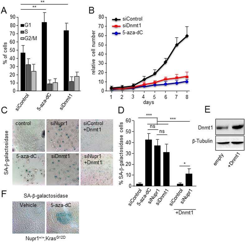
Figure 3. DNA hypomethylation triggers OIS in pancreatic cancer cells.
(A) Arrest in cell cycle and accumulation of G1 cells is observed in 5-aza-dC and siDnmt1 treated cells. (B) Proliferation curve showing that either 5-aza-dC treatment or siRNA-mediated Dnmt1 silencing stops cell proliferation. The relative cell number at each time point on the growth curves represents the means value ± SD of triplicates normalized to the cell number at day 1. (C) SA-βGal activity staining in MiaPaCa2 cells. As with siNupr1 treatment (upper middle panel), 5-aza-dC (lower left panel) and siDnmt1 (lower middle panel) treatments induce cells enter in senescence. On the other hand, the constitutive overexpression of Dnmt1 in MiaPaCa2 cells significantly rescues the siNupr1-induced senescence (lower right panel and right plot). (D) quantification of SA-βGal staining. (E) Expression of DNMT1 in MiaPaCa2 transfected with empty and pCMV6-Dnmt1 plasmid (F) SA-βGal staining in pancreas from Nupr1+/+ KrasG12D-expressing animals treated with 5-aza-dC (n = 6) or vehicle (n = 6). Means ± SD; ns = no significant, *p < 0.05, **p < 0.01, ***p < 0.001. Scale bar, 20 μm.

