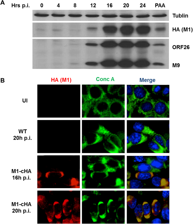
Figure 3. M1 localizes to cellular ER during MHV-68 infection.
(A) NIH3T3 cells were infected with M1-cHA MHV-68 at MOI 10 and harvested at indicated time points for western blot analysis using antibodies specific for tublin (top), HA (middle), ORF26 and M9 (bottom). (B) NIH3T3 cells were mock-infected or infected with wild-type (WT) or M1-cHA MHV-68 at MOI 10 and fixed for IFA analysis as described in Materials and Methods using antibodies against HA (red), concanavaline A (conc A) (green) and Hoechst (Blue).

