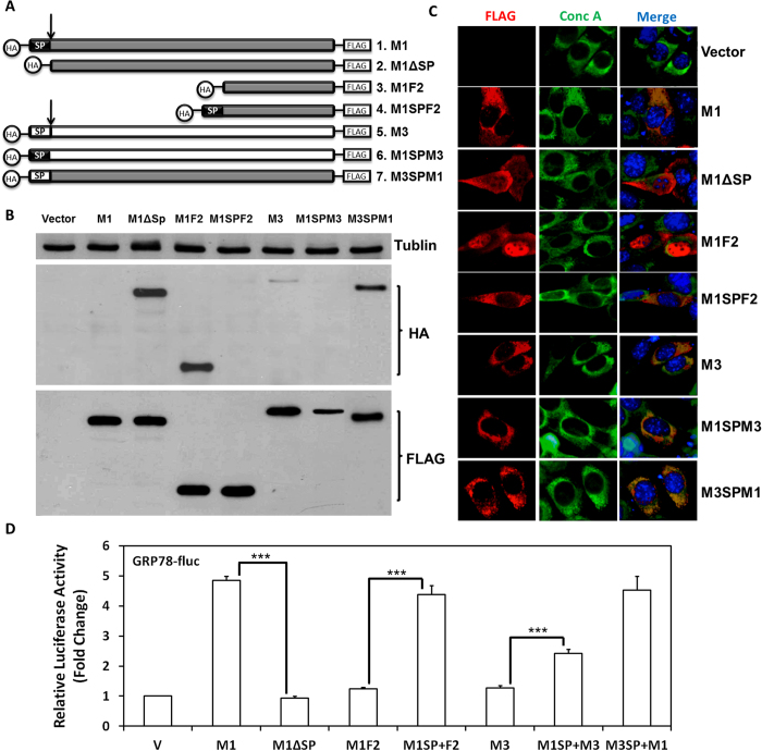
Figure 4. M1 requires ER localization for ER chaperone induction.
(A) Schematic diagram of the M1 mutant constructs. (B) 293T cells were transfected for 24 hours with the plasmids encoding the indicated M1 mutants and harvested for western blot analysis using antibodies against tublin (top), HA (middle) or FLAG (bottom). (C) NIH3T3 cells transfected with the indicated M1 mutant constructs were fixed and analyzed by IFA using antibodies specific for FLAG (red), conc A (green) and Hoechst (Blue). (D) 293T cells were transfected with the GRP78-fluc, PGK-renilla-luciferase, and plasmids encoding each indicated M1 mutants. Cell lysates were collected 24 hours posttransfection and were analyzed by dual-luciferase assays as described in Fig. 1A.

