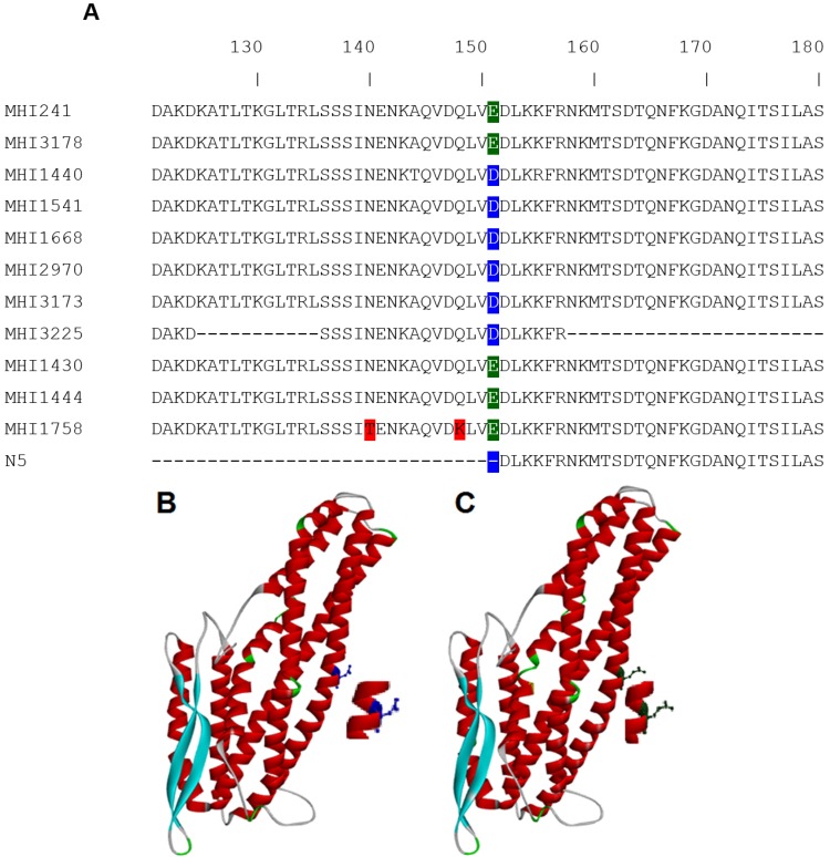
Figure 1.
(A) Partial NheB sequences comprising amino acid 121–180. The complete Clustal Omega alignment file is given as Figure S1. Although several primers were applied, sequencing of MHI 3225 was only partially possible for unknown reasons. The most prominent difference is the point mutation at position E151D compared to the reference strain MHI 241. This mutation is not present in a formerly-published rNheB fragment, which always tested negative in EIA and Western blot; (B) position of the aspartic acid residue in the structural model of NheB; and (C) the glutamic acid residue is protuding more and slightly rotated in the model.

