Figure 6.
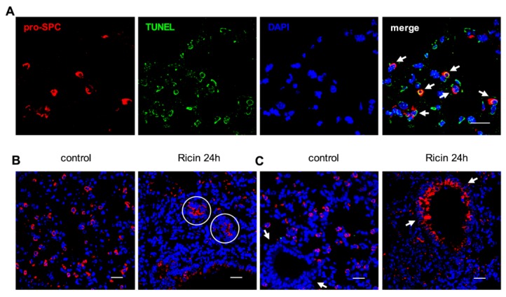
Immunofluorescence analysis of ATII cells after ricin exposure. (A) Co-immunofluorescence for pro-SPC (red), TUNEL (green) and DAPI (blue) from lung tissue 24 h after intoxication. Arrows point to TUNEL-positive, apoptotic ATII cells; (B) Immunofluorescence for pro-SPC (red) and DAPI (blue) staining of lung tissue from naive (left) and 24 h post-intoxicated (right) mice. Circles indicate pro-SPC+ ring structures; (C) Visualization of bronchiole structures (arrows) stained for pro-SPC (red) and DAPI (blue) in lung tissue from naive (left) and 24 h post-intoxicated (right) mice. Scale bars indicate 20 µm.

