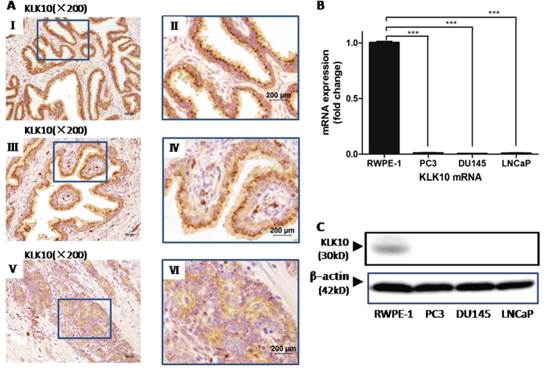
Figure 1. Expression of KLK10 is low in prostate cancer tissue and cell lines.
(A: I, II) KLK10 protein grains presented brown and were regularly arranged in the cytoplasm of BPH tissue. (A: III, IV) KLK10 protein gains presented brown and were regularly arranged in the cytoplasm of matched adjacent normal tissue of prostate cancer. (A: V, VI) KLK10 protein grains were lighter or even absent and were irregularly arranged in the cytoplasm of prostate cancer tissue. (B) KLK10 mRNA expression was significantly lower in PC3, DU145 and LNCaP clone FGC than in RWPE-1. (C) KLK10 protein expression was almost lost in PC3, DU145 and LNCaP clone FGC compared with that in RWPE-1.

