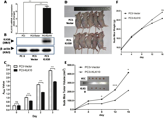
Figure 2. KLK10 gene inhibits the proliferation of PC3 cells.
(A,B) After transducing the KLK10 gene into the PC3 cell line, the mRNA and protein expression levels of KLK10 were significantly higher in PC3-KLK10 than in PC-3 and the blank Vector group. (C) CCK-8 cell proliferation assay showing the growth deceleration of PC3-KLK10 cell lines 24 h after cell seeding, and this phenomenon lasted for 3 days (P < 0.001). (D) General images of PC3-KLK10- and Vector-transplanted tumours showed that the tumour volume was smaller in the KLK10-over-expressing group than in the Vector group. (E) Tumour volume line graph showed that tumours were successfully generated in nude mice 4 days after cell transplantation; the PC3-KLK10 tumour grew more slowly than the PC3-Vector tumour, and a significant difference in tumour volume was found between these two groups starting from the eighth day. (F) Weight line graph showed that the mouse weight was lower in the PC3-Vector group than in the PC3-KLK10 group from the eighth day, but the difference was not significant (P > 0.05).

