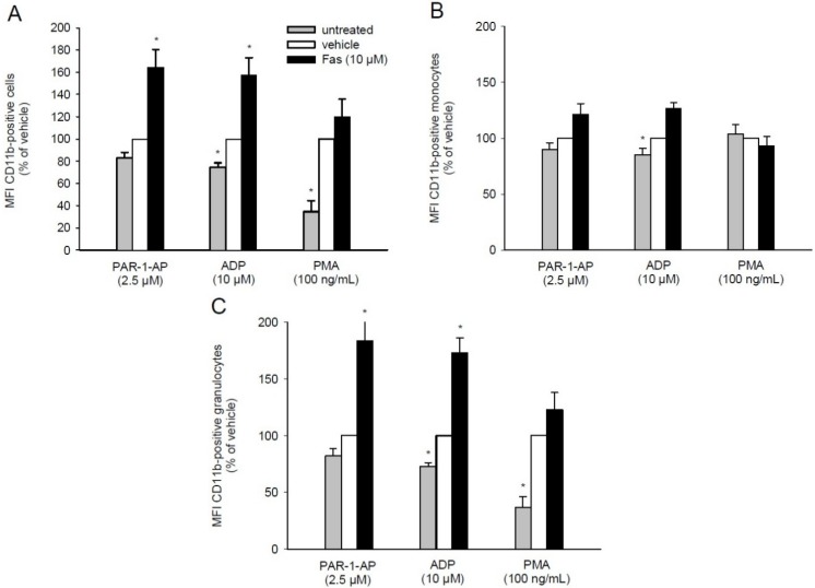
Figure 5.
Effect of fascaplysin on leukocytic CD11b expression. (A–C) Whole blood was incubated with 10 μM fascaplysin (black bars, n = 6) or vehicle (DMSO, white bars, n = 6) for 0.5 h followed by stimulation with PAR-1-AP, ADP or PMA. Data are given in % of vehicle. Unstimulated whole blood served as negative control (grey bars, n = 6). Expression of CD11b on all leukocytes (A), monocytes (B) and granulocytes (C) were assessed by flow cytometry using double fluorescence staining (CD45/CD11b). Mean ± SEM. * p < 0.05 vs. vehicle.

