Figure 6.
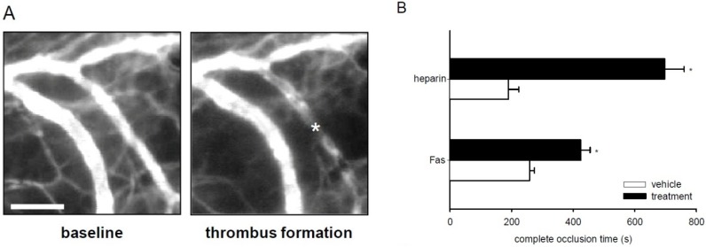
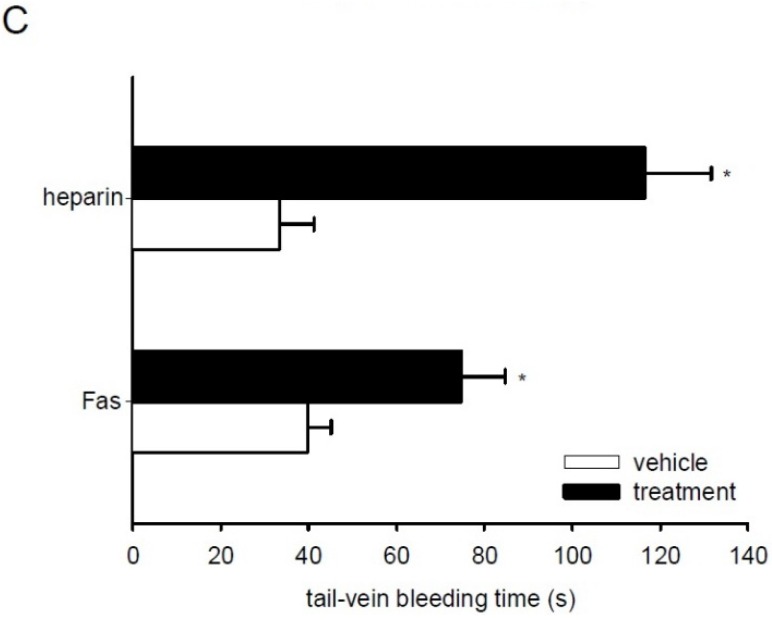
Effect of fascaplysin and heparin on thrombus formation and tail-vein bleeding time. (A) Intravital fluorescent microscopic images of a postcapillary venule in the dorsal skinfold chamber of a vehicle-treated BALB/c mouse before (baseline) and after photochemically induced thrombus formation (asterisk). Blue-light epi-illumination with contrast enhancement by 5% fluorescein isothiocyanate (FITC)-labeled dextran 150,000. Scale bar: 50 μm. (B) Complete occlusion time of postcapillary and collecting venules upon photochemically induced thrombus formation in dorsal skinfold chambers of heparin-treated (upper black bar, n = 3), fascaplysin-treated (lower black bar, n = 6), and vehicle-treated BALB/c mice (upper white bar, saline, n = 3 and lower white bar, DMSO, n = 6), as assessed by intravital fluorescence microscopy. Mean ± SEM. * p < 0.05 vs. vehicle. (C) Tail-vein bleeding time of heparin-treated (upper black bar, n = 3), fascaplysin-treated (lower black bar, n = 6), and vehicle-treated BALB/c mice (upper white bar, saline, n = 3 and lower white bar, DMSO, n = 6). Mean ± SEM. * p < 0.05 vs. vehicle.


