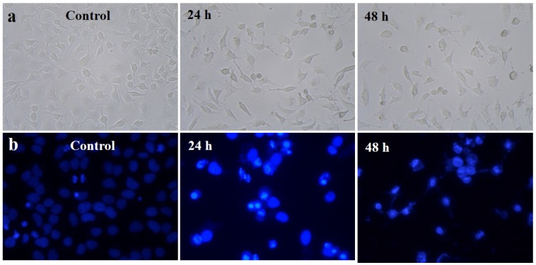
Figure 7.
(a) Phase contrast microscopic images of gold nanoparticles induced gross cytomorphological changes and growth inhibition at different time (24 h and 48 h) intervals on the HeLa cells; (b) 4′,6-diamidino-2-phenylindole dihydrochloride (DAPI) staining shows apoptotic and necrotic cell death due to the cytotoxicity of biosynthesized gold nanoparticles (24 h and 48 h).

