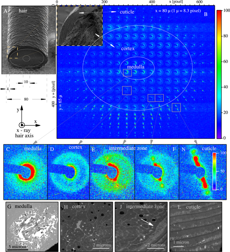
Figure 1.
SEM image of a human hair fiber is shown in (A) with schematic overlays on to the cut face of the hair fiber to emphasize the relative size of the hair and the main hair regions. In (B), we spatially position the SAXS diffraction patterns relative to each other, so that the locations match the position of the x-ray beam on the sample, thus generating a SAXS “map” of human hair. This map allows us to see the spatial variation of the SAXS patterns from a 30-micron- thick cross-section of hair. The incident x-ray beam is  to the hair axis. SEM zoom of selected cross section from (A) is overlapped to the SAXS map showing the real-space cross section of the cuticle and cortex. Magnified views of the characteristic SAXS patterns are shown in (C) for the medulla, (D) for the cortex, and in (N) for the cuticle. (E,F) are for the newly discovered intermediate zone. Corresponding Scanning Transmission Electron Microscopy (STEM) images shown in (G,H,J,L).

