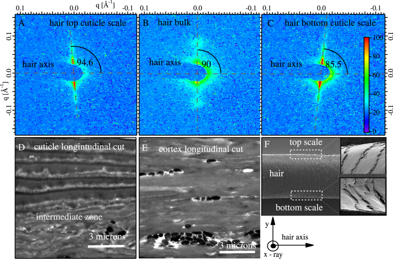
Figure 3.
The SAXS patterns taken on a single hair with the incident X-ray beam ⊥ to the hair axis is shown in (A) to (C). (A) is for the top of the hair in the cuticle, (B) is largely the cortex region and (C) for the bottom edge of the hair in the cuticle region. Corresponding real- space STEM images of hair cut longitudinally are shown in (D) (cuticle) and (E) (cortex) with no staining or coating. In (D), we again observe the intermediate zone. (F) shows SEM images of the hair and the detail of the cuticle shows that the’s scales are oriented with an angular offset with respect to the hair’s axis. The angular offset between the hair’s axis and the cuticle scales in (F) is the same angular offset we observe in reciprocal space between (A,B), or (B,C).

