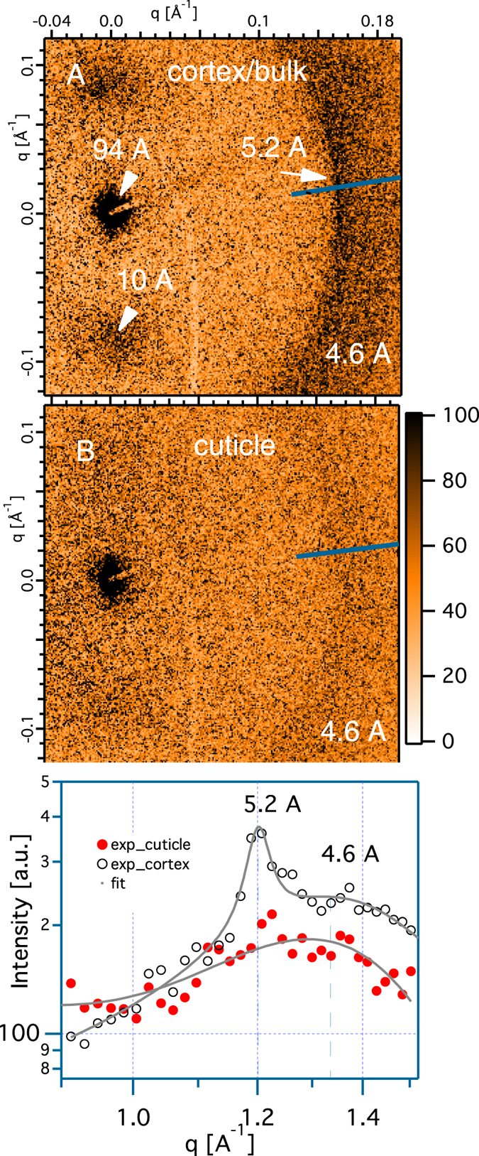
Figure 4.

Comparison of the wide angle x-ray scattering of cortex shown in (A) and cuticle shown in (B) measured with x -ray beam ⊥ to the hair’s axis. (C) showing the line projections of the integrated intensities for the peak 5.2 Å and the peak 4.6 Å of the two respective regions.
