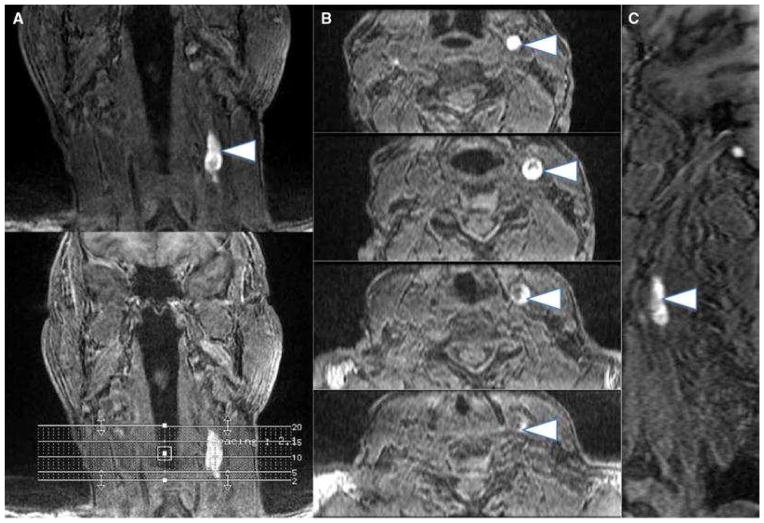
Figure 2.
Multiplanar images (a coronal, b axial, c sagittal) of carotid intraplaque hemorrhage (arrowheads) within the wall of the left carotid artery. Intraplaque hemorrhage is detected using a 3D-T1-weighted fat-suppressed fast field echo sequence by exploiting the T1 shortening effects of methemoglobin and the technique has been histologically validated
From Singh N, Moody AR, Rochon-Terry G, Kiss A, Zavodni A. Identifying a high risk cardiovascular phenotype by carotid MRI-depicted intraplaque hemorrhage. Int J Cardiovasc Imaging. 2013 Oct;29(7):1477–83; with permission.

