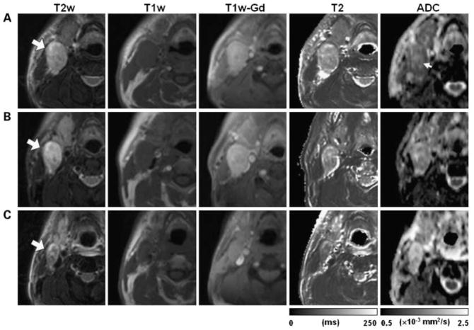
Figure 1.
Array of representative images of a HNSCC patient with partial response after treatment. Images in each row are from three measurement time points: pretreatment (A), 1 week after radiation therapy (B), and 2 weeks after the completion of the treatment (C). Large arrows, same metastatic nodal mass that was followed through the treatment course; small arrow, central region of the mass with ADC higher than the rim. (From Kim S, Loevner L, Quon H, et al. Diffusion-weighted magnetic resonance imaging for predicting and detecting early response to chemoradiation therapy of squamous cell carcinomas of the head and neck. Clinical cancer research : an official journal of the American Association for Cancer Research 2009;15:986–94, with permission.)

