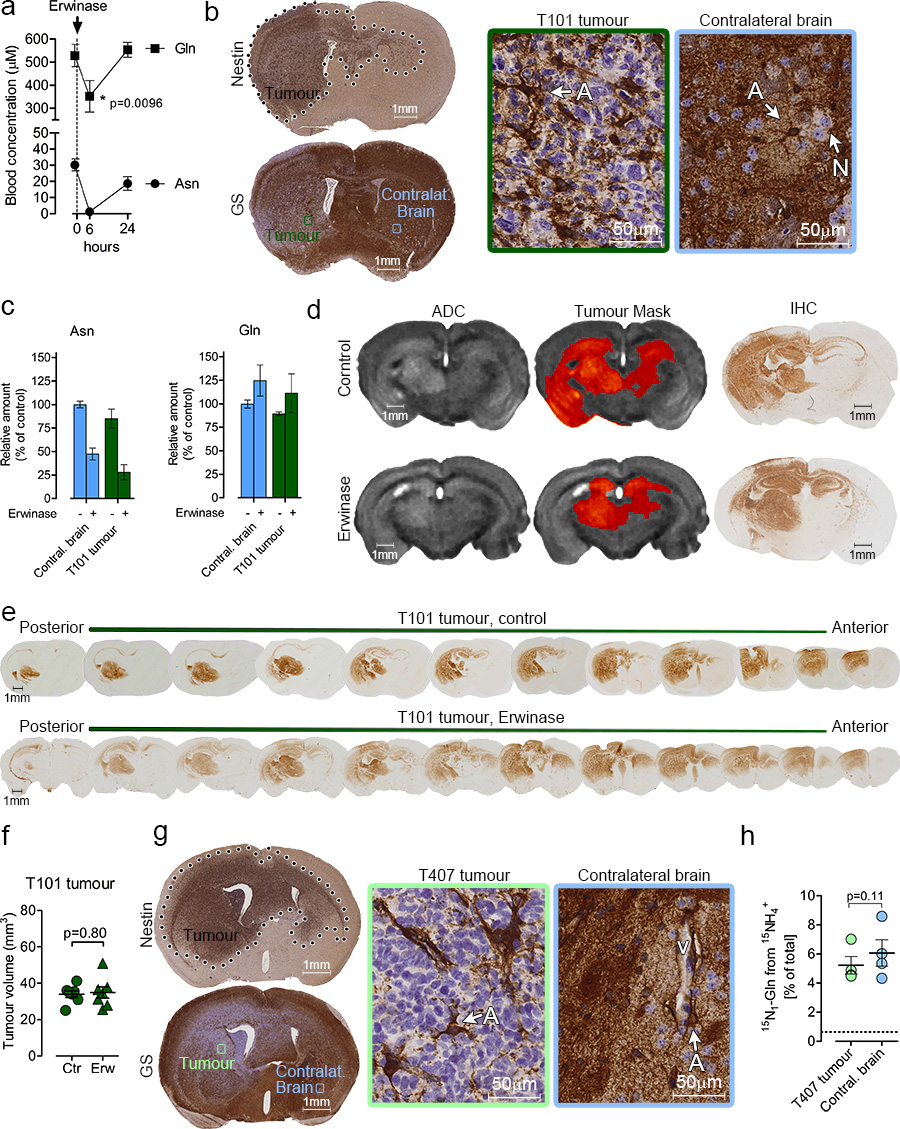
Figure 7.
Glutamine supply for GBM tumours with low GS expression. (a) Gln and Asn levels were measured by HPLC-MS in peripheral blood samples obtained at indicated time points from immunocompromised mice intraperitoneally injected with Erwinase (5U/gr of body weight). Mean ± S.E.M. n=5 mice. p values refer to a two-tailed t test for paired samples. (b) Coronal section of human T101 GBM xenograft grown in brain of immunocompromised mice, and stained for human nestin and GS. Left panels are magnification of the respective framed regions. A: astrocytes, N: neuron. (c) Immunocompromised mice were orthotopically implanted with T101 GBM tumours and treated with Erwinase for 6 weeks as described in the Methods section. Asn and Gln were assessed in the tumour and contralateral brain 6h after the last Erwinase injection. Mean ± S.E.M. n=3 mice. (d) MRI-based Apparent Diffusion Coefficient (ADC) maps of T101 GBM tumours treated with Erwinase as described in (c). The tumour mask has been manually delineated to highlight the tumour region. IHC staining of brain sections corresponding to the MRI scans are shown. T101 tumours were stained with an anti-human EGFR antibody. (e) Two representative series of coronal sections of T101 brain xenografts, were stained for human EGFR. Mice were treated with Erwinase as in (c). (f) Volumes of T101 orthotopic tumours obtained thorough quantitative imaging of EGFR-stained serial sections of brains. Mice were treated with Erwinase as in (c). Mean ± S.E.M. n=7 mice. p value refer to a two-tailed t test for unpaired samples. (g) Coronal section of human T407 GBM xenograft grown in brain of immunocompromised mice, and stained for human nestin and GS. Left panels are magnification of the respective framed regions. A: astrocytes, V: blood vessel. (h) 15N1-Gln enrichment in T407 GBM tumours and in contralateral brains, after a 4h intracarotid infusion with . The dashed lines correspond to the natural abundance of 15N1-Gln. Mean ± S.E.M. n=4 mice. p value refer to a two-tailed t test for paired samples.

