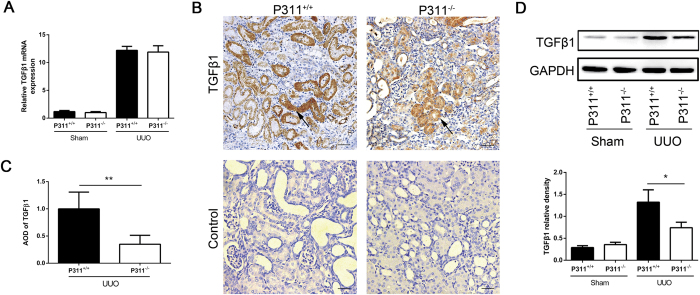
Figure 5. P311 deficiency suppresses TGF-β1 expression in UUO mice.
(A) RNA was isolated in kidneys from P311+/+ (n = 6) and P311−/− (n = 6) mice 7 days after UUO or sham operation. TGF-β1 mRNA expression was determined by real-time PCR. (B) Representative photomicrographs of TGF-β1-specific immunohistochemical staining of UUO kidneys from P311+/+ and P311−/−mice on day 7. Black arrowhead indicates the TGF-β1-positive region. Bottom panel: negative controls for the immunohistochemical staining of TGF-β1 on the obstructed kidneys from both P311+/+ and P311−/− mice. (C) TGF-β1-positive region was quantified in stained sections from P311+/+ (n = 6) and P311−/− (n = 6) mice 7 days after UUO. (D) Western blot analysis of TGF-β1 protein levels. TGF-β1 protein levels in each treatment group (n = 3 per group) were quantified. A-D are representative of at least three similar experiments. Scale bar: 100μm. Data are presented as the mean ± SD. *P < 0.05; **P < 0.01.

