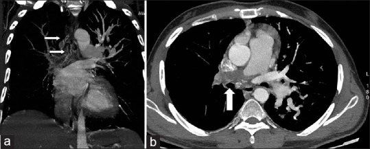
Figure 1.

(a, b) Coronal maximum intensity projection (MIP) images of CT bronchial angiography showing hypertrophied right bronchial arteries (arrows in a). One of the bronchial arteries was arising from the intercostobronchial trunk from the descending aorta (not shown here). Origin of the second right bronchial artery was, however, not clear on CT angiography. A thrombus was seen in the right pulmonary artery (arrow in b) extending till the subsegmental branches
